Salivary Gland Pathologies
Salivary gland pathologies: clinical cases, treatments and procedures.
Below are a few video cases related to salivary gland conditions.
Sialorrhea
Excess saliva production from one or more salivary glands.
Sublingual gland infection
Infection of the sublingual gland. You can observe pus drainage.
Duct patency and drainage
Patency and drainage of the sublingual duct.
Parotid Adenoma
This is a benign disease in the parotid gland (a salivary gland), which nonetheless requires removal.
Pleomorphic adenoma of the parotid gland
Pleomorphic adenoma is a benign pathology of the salivary glands.
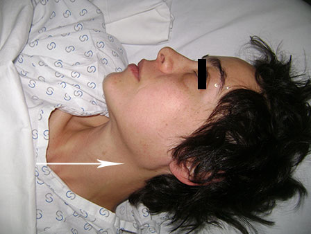
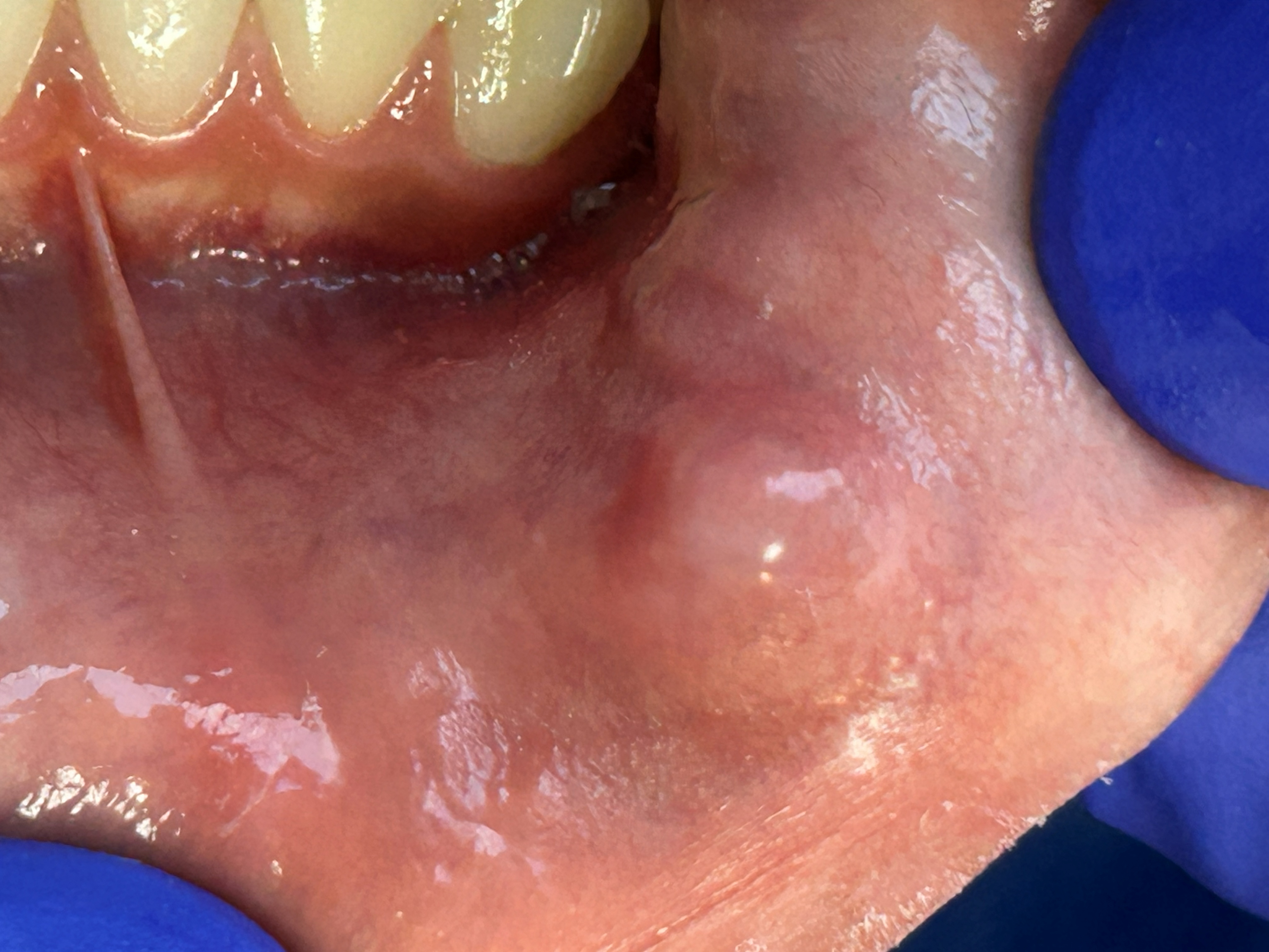
Submaxillary Adenoma
Stones (calcium deposits) in the parotid gland
This is a condition which can occur in the salivary glands consisting of multiple calcium deposits, forming stones within a gland or duct. These deposits necessarily require surgical removal.

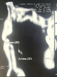
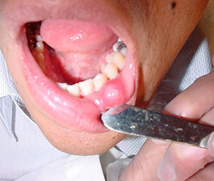
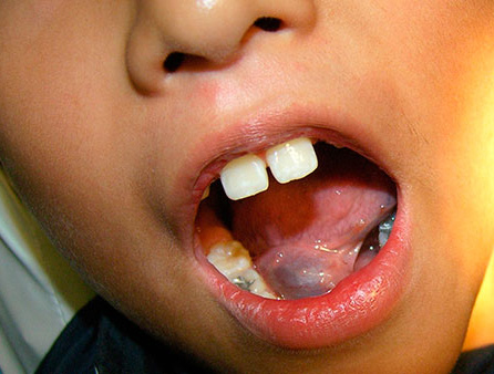
Lingual Sialolith
Sialoliths are stones formed by calcium deposits in the ducts and salivary glands.
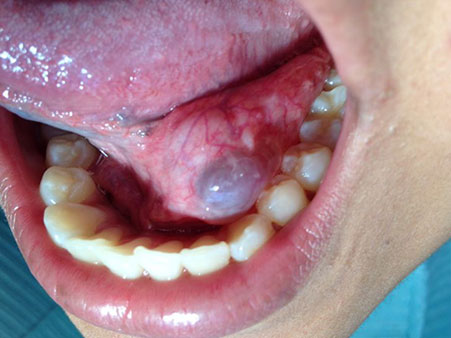
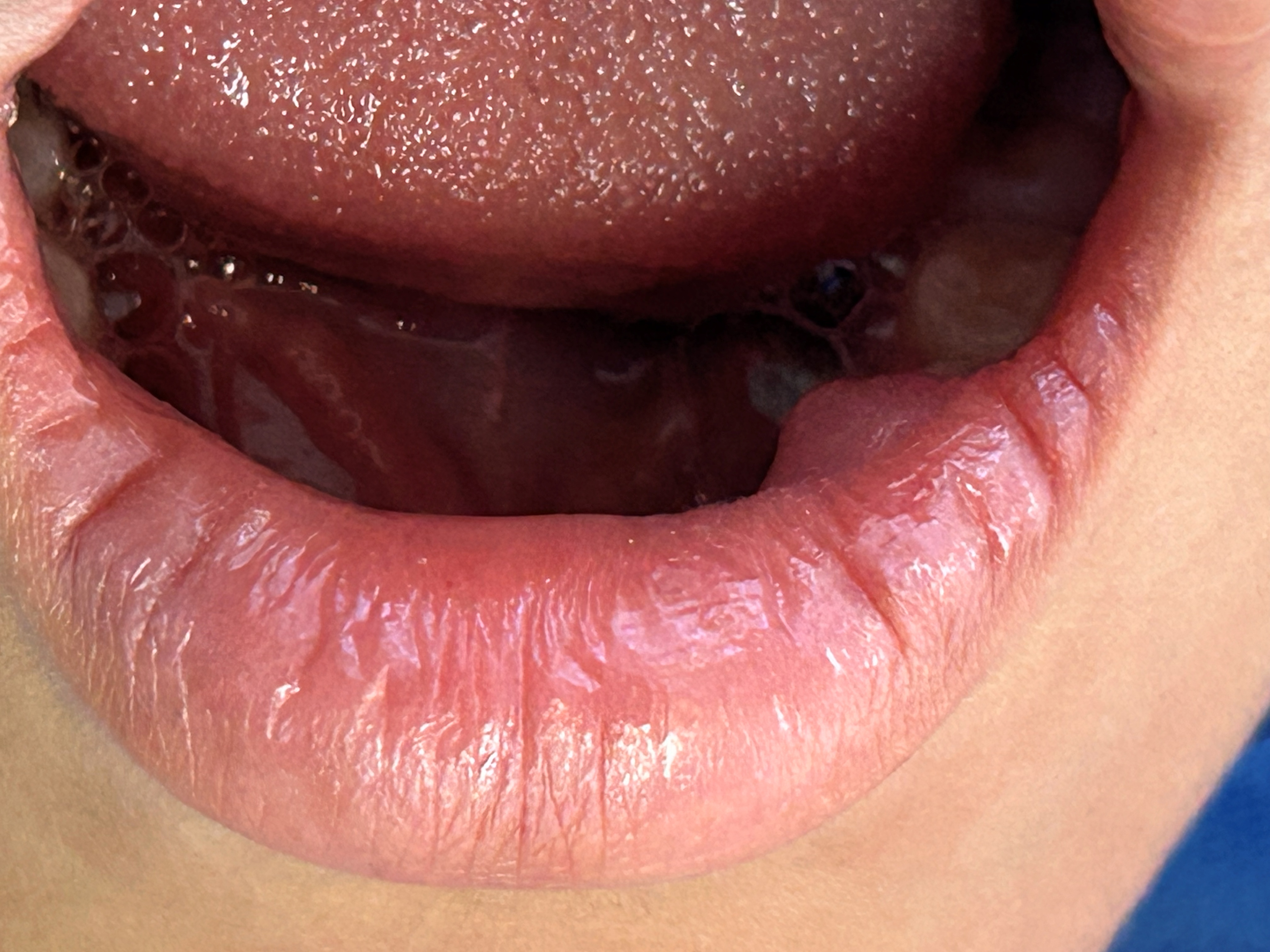
Mucocele
Mucoceles are salivary retention cysts that are usually formed by a direct traumatic event to the salivary gland. These cysts can increase in size as they fill with saliva. These cysts also require surgical removal.
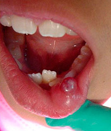
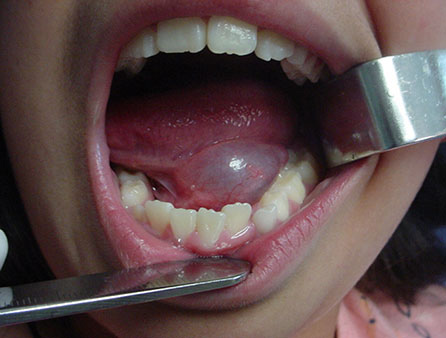
Ranula
A ranula is a salivary retention cyst on the floor of the mouth that also requires surgical removal.
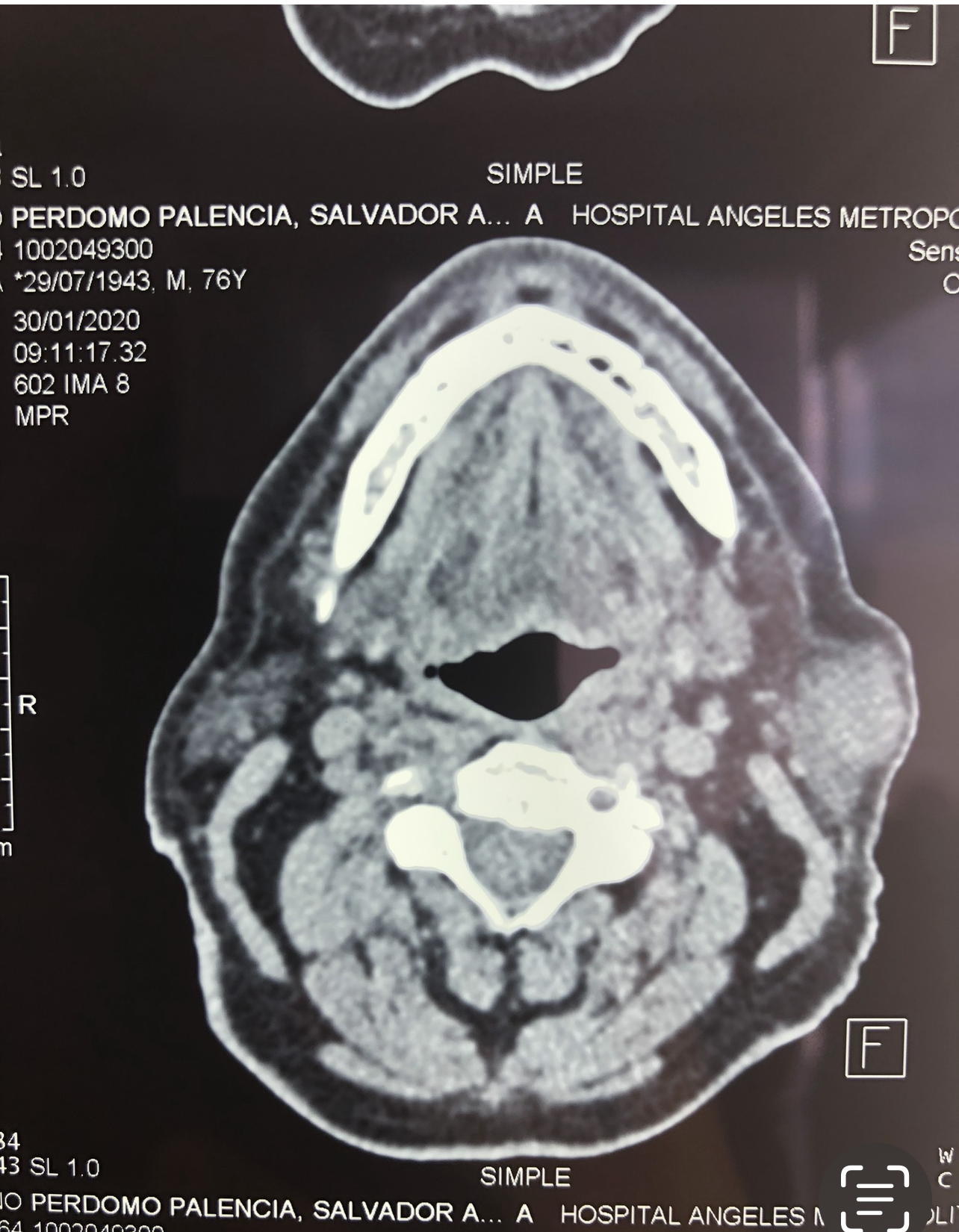
Parotid gland cyst
We can also find cysts that have formed in the major salivary glands as shown in the figure below.
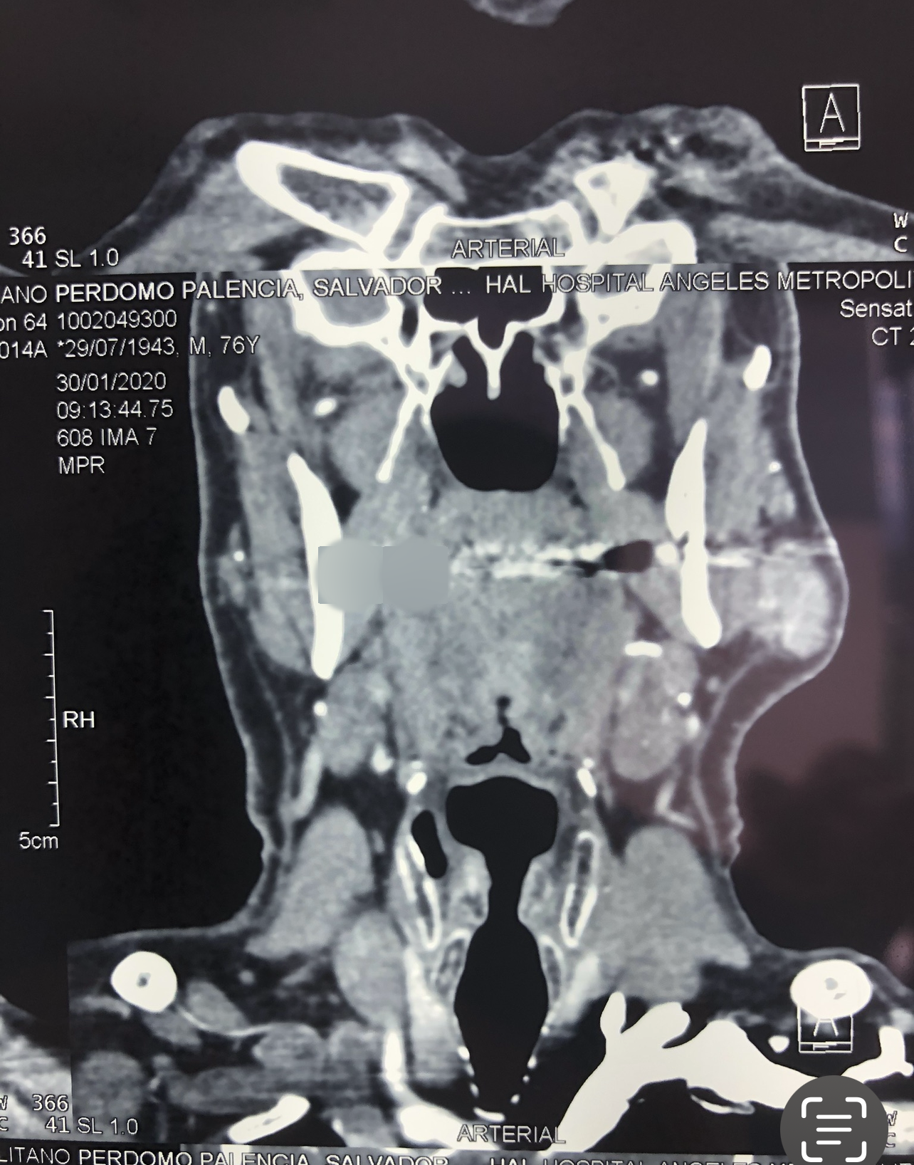
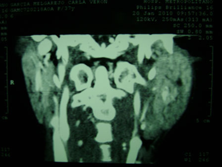
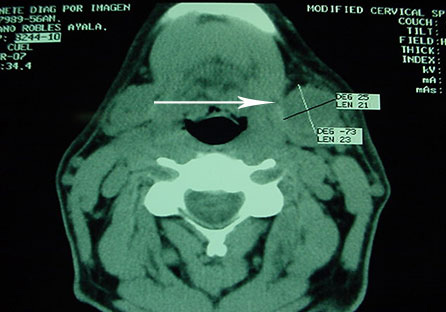
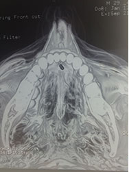
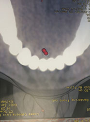
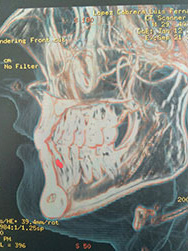
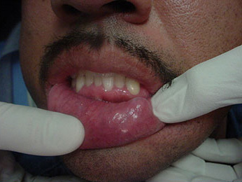
Sialolith in the Submandibular Gland
Sialoliths are calcium deposits (stones) that can form within salivary ducts or glands. Depending on size and location, surgical removal may be recommended.
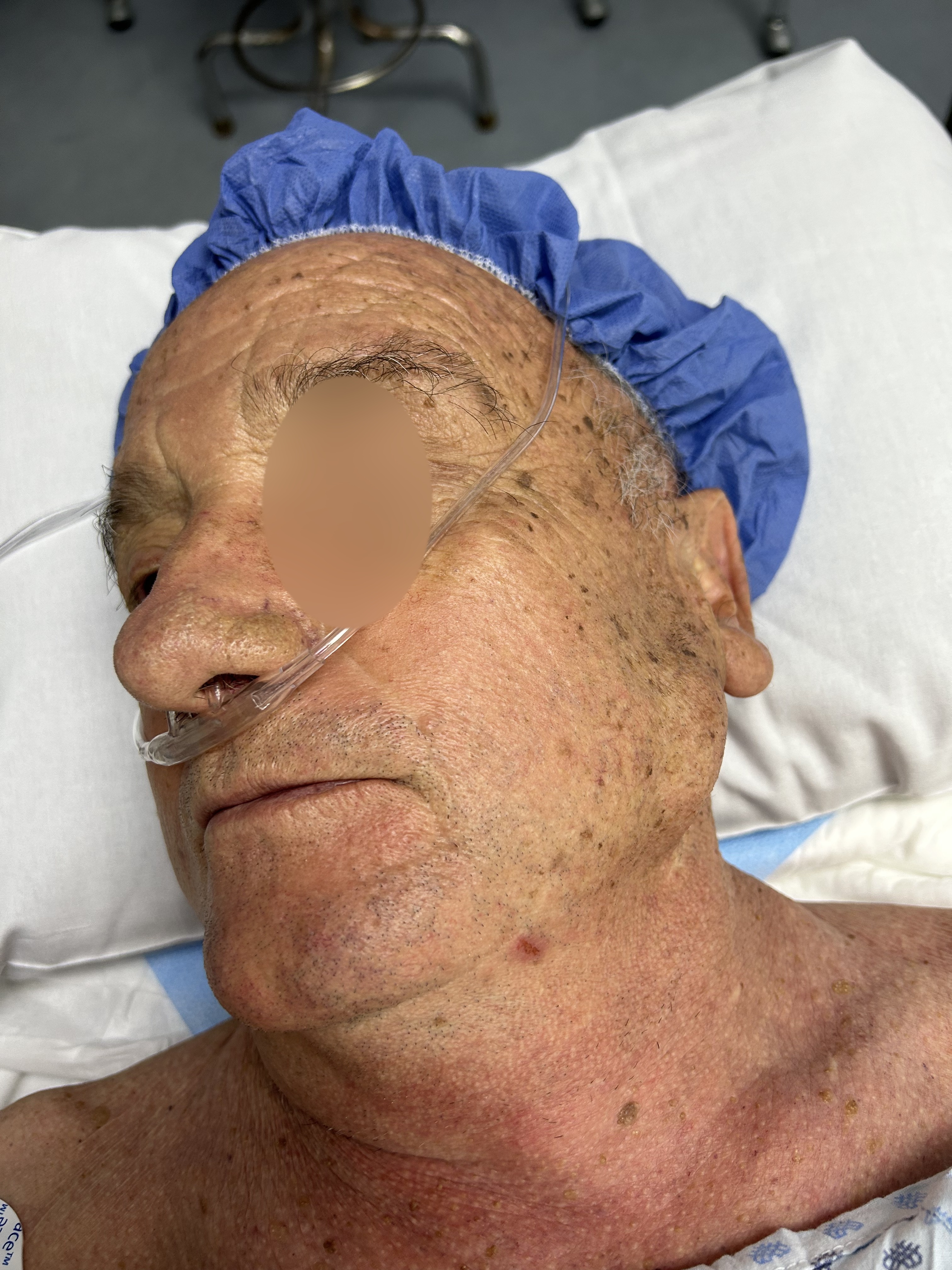
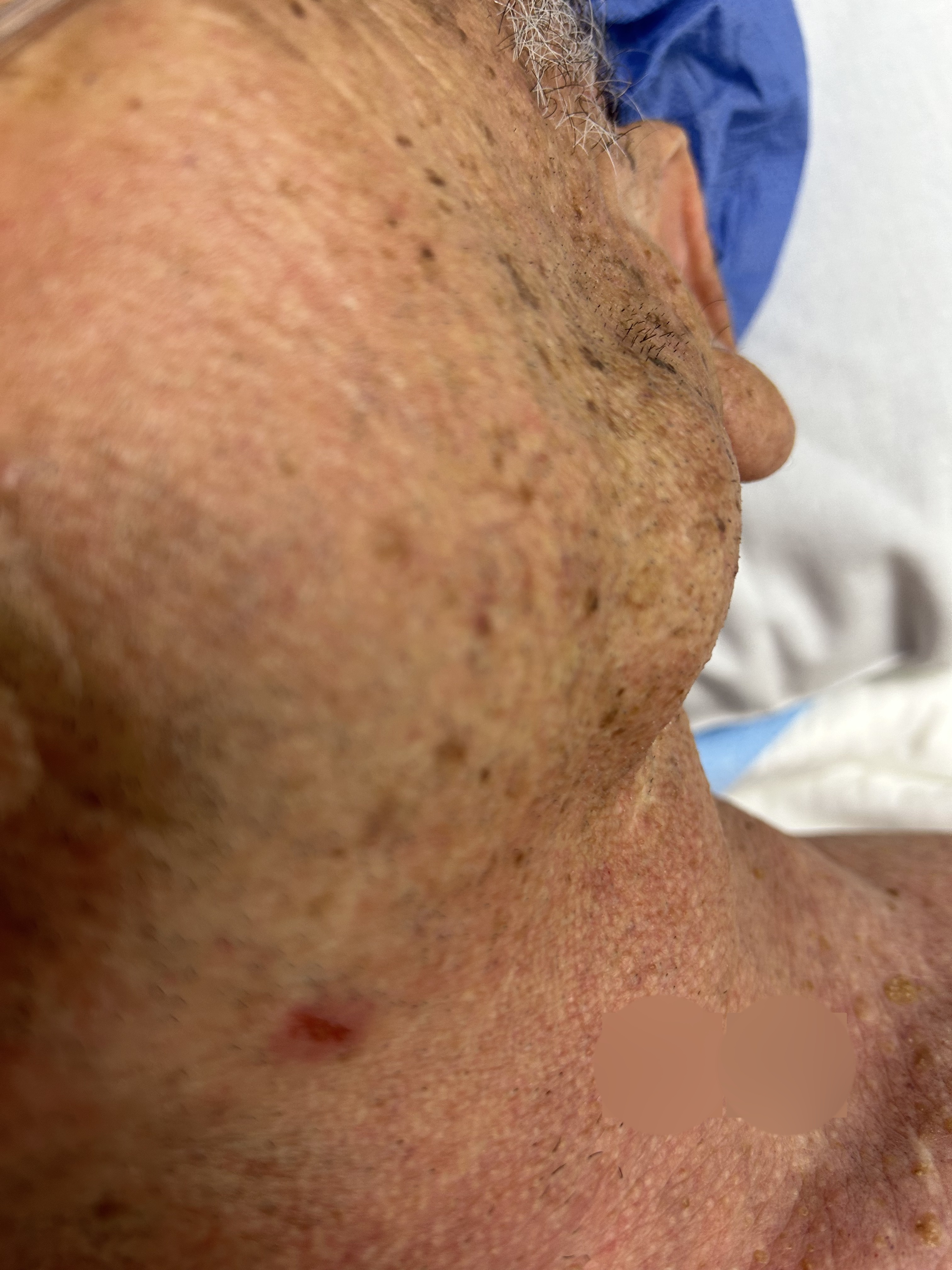
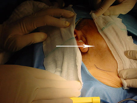
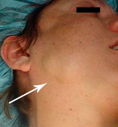
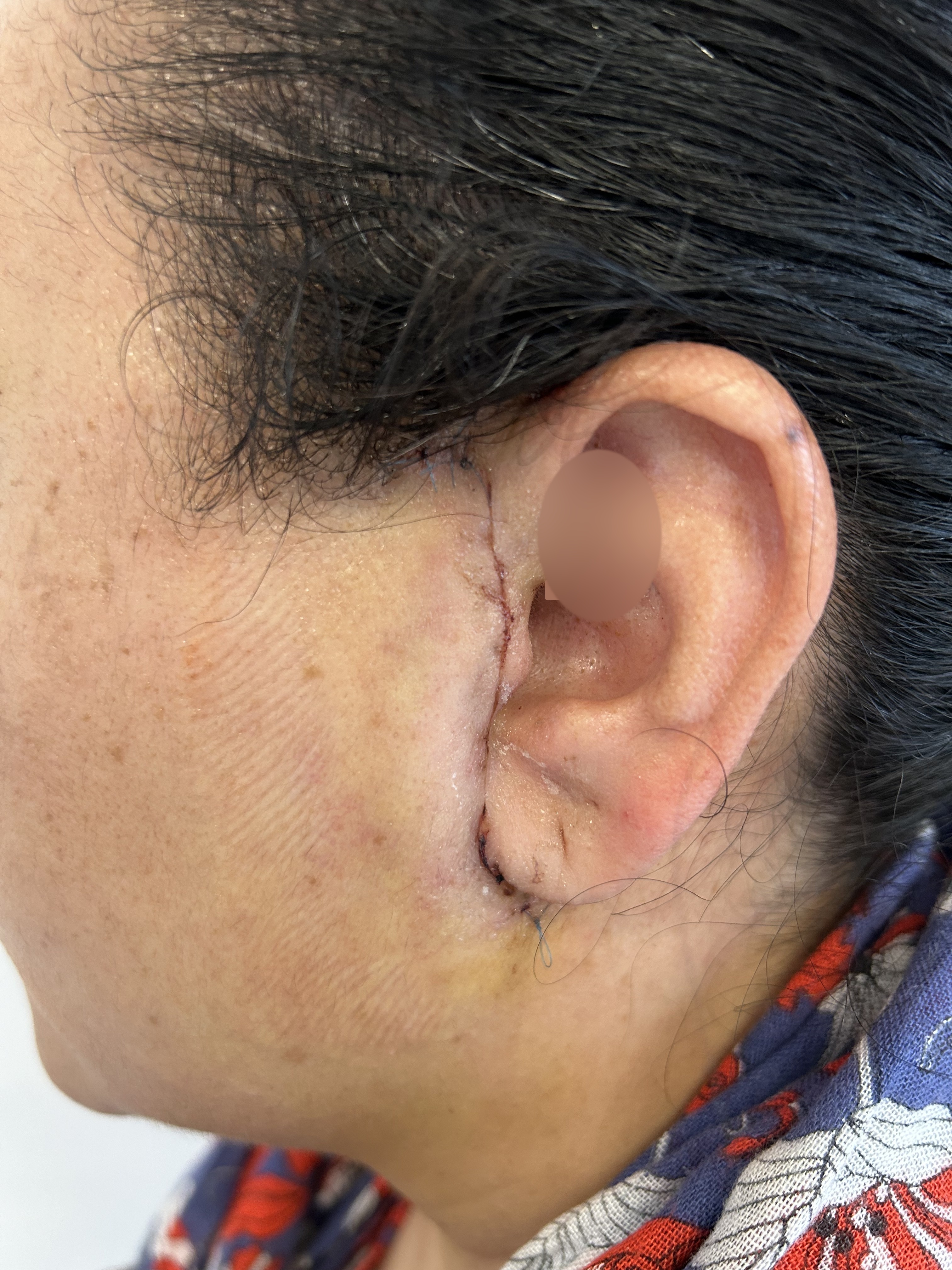
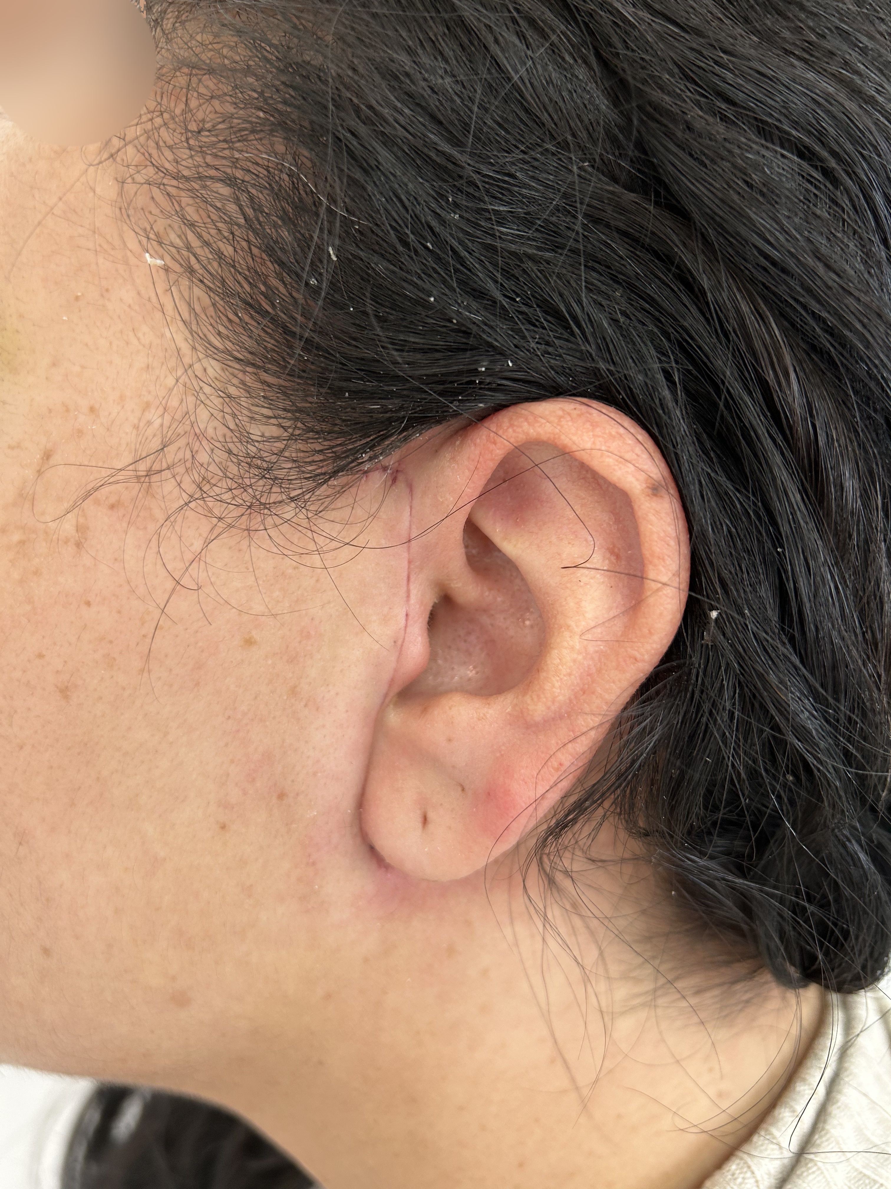
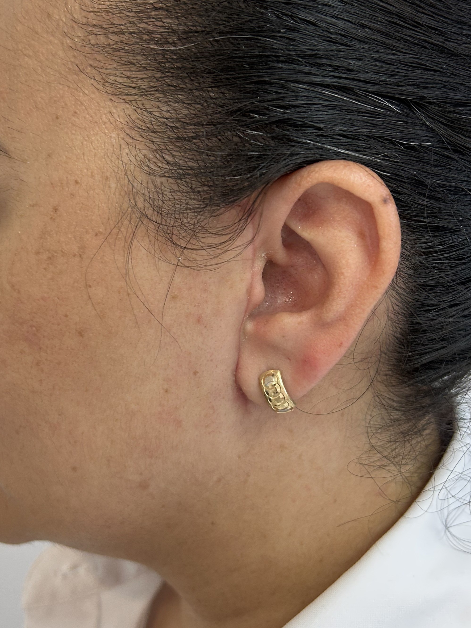
Case: Adenoma — follow-up of the surgical approach (scar)
Clinical case of an adenoma in a salivary gland. Clinical assessment and imaging help define the surgical approach and follow‑up plan.
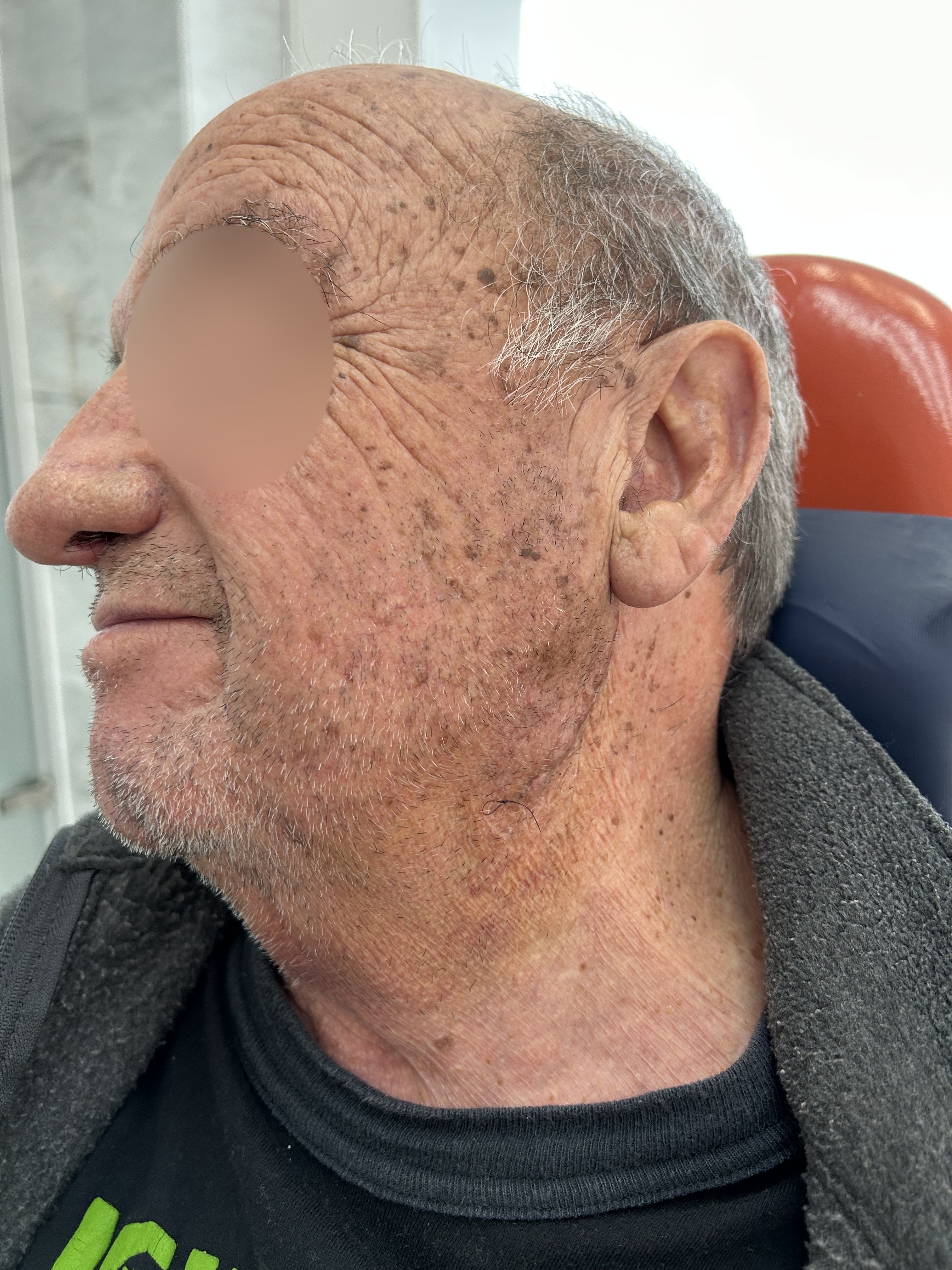
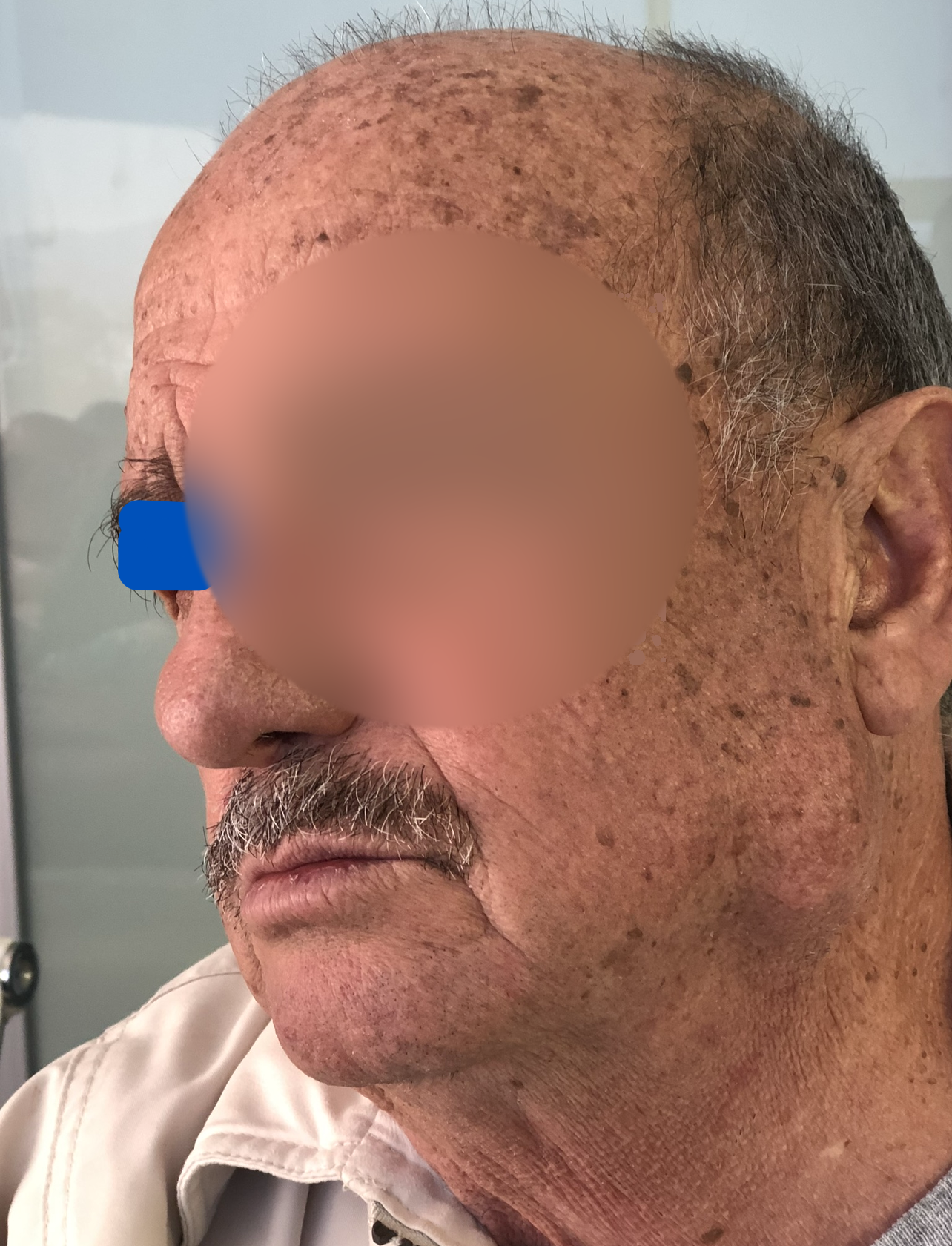
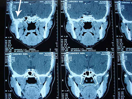
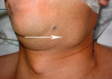
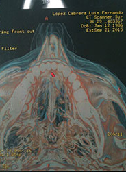
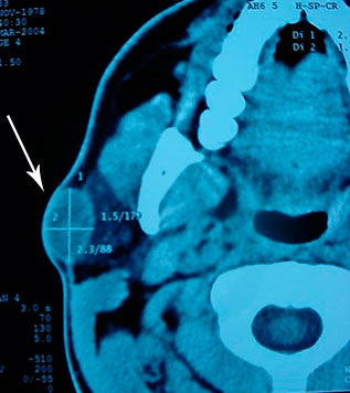
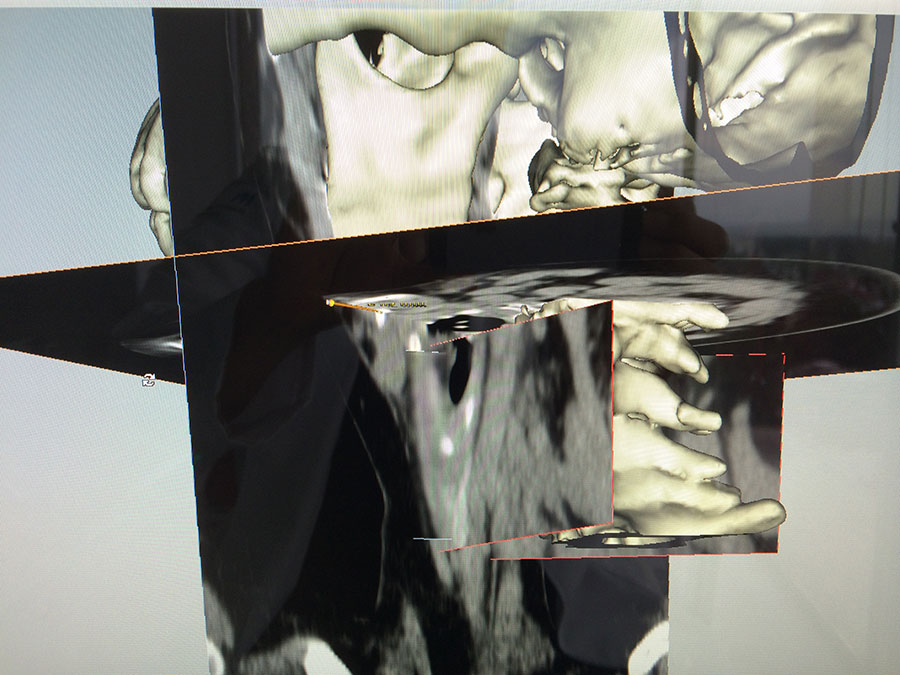
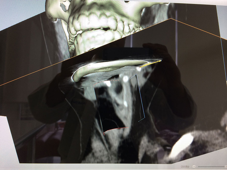
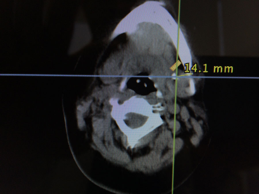
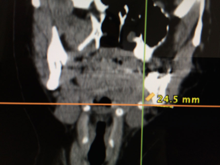
Case: Parotid Tumor
Clinical case of a parotid gland tumor. Depending on location, size and diagnosis, it may require surgical resection and postoperative follow‑up.